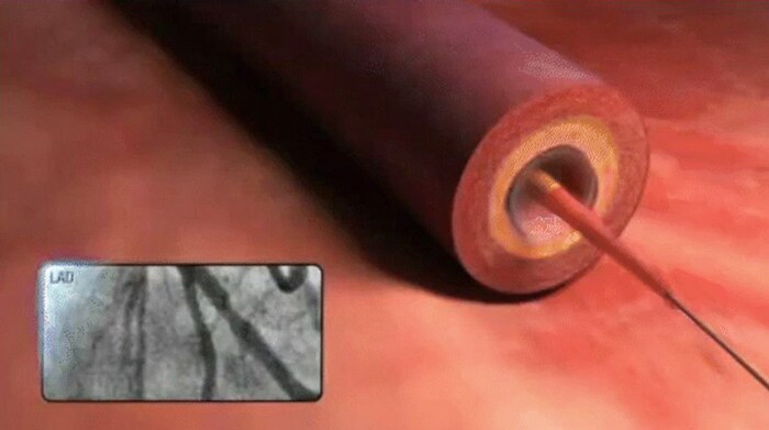
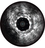
Intravascular ultrasound (IVUS) is a catheter-based imaging technology that allows physicians to visualize blood vessels from the inside out. Cross-sectional images help assess presence and extent of disease, plaque geometry and morphology, guide wire position during lesion crossing, and stent position post-treatment. The imaging transducer emits high-frequency sound waves that echo off vessel walls and are sent back to the system in varying intensities depending on the tissue. System electronics process the signal to display the cross-sectional image.
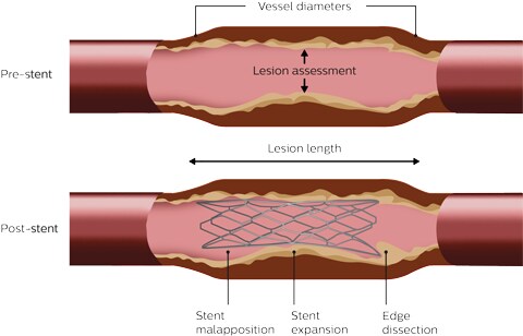
Plan the procedure with pre-stent IVUS to size stents and identify the optimal landing zone. Perform post-stent IVUS to confirm good stent expansion, apposition and no edge dissection.
Landing zone/stent length
Lesion
Assess lesion characteristics to guide plaque modification strategy.
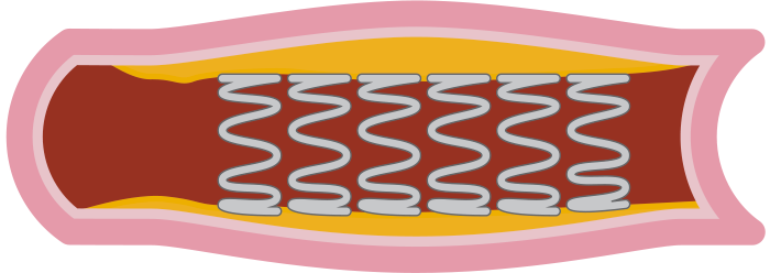
Land and expandPost-stent IVUS to guide and confirm stent optimization
Landing zone
Expansion and apposition
MSA ≥90% of the distal reference lumen area and full stent apposition throughout.
Stent edges
No edge dissection involving media with length > 3mm and arc ≥ 60°
Criteria used for IMPact on Revascularization Outcomes of intraVascular Ultrasound Guided Treatment of Complex Lesions and Economic Impact (IMPROVE) trial. Shlofmitz et al. 1
Land and expand

The ADAPT-DES study reported that IVUS guidance was associated with a change in PCI strategy 74% of the time. 2 Most often the impact was a larger size of stent or balloon, followed by post-dilation. Furthermore, the study reported that larger stent areas resulted in cases where both pre- and post- PCI IVUS were performed compared to when only post-PCI IVUS was performed. 2
Easily assess lesion characteristics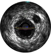
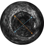
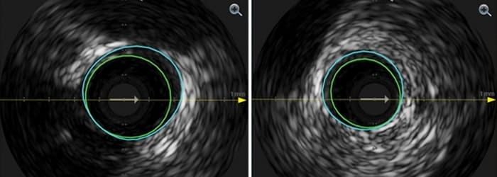
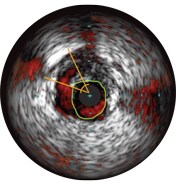
Vessel diameters may be determined at proximal and distal reference sites by obtaining lumen diameters, mid-wall diameters (halfway between lumen and vessel), or vessel diameters, in order of increasing aggressiveness.
If maximum and minimum diameters are used, measurements should bisect the geometric center of the vessel rather than the center of the IVUS catheter.
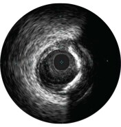
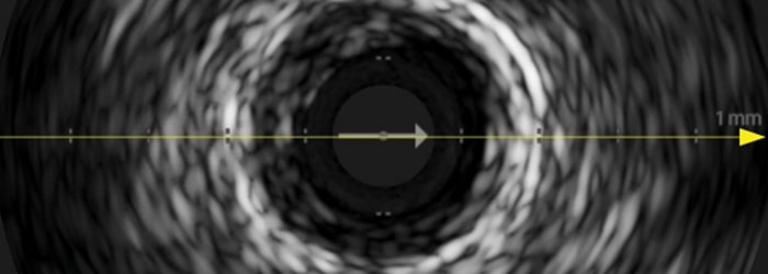
IVUS can help clarify degree and type of stenosis (i.e, MLA, plaque burden, and calcium). While IVUS can also characterize plaque rupture, thrombus, and dissection, calcium may be more common in everyday PCI. An important factor in your stenting strategy, calcium is characterized by very bright areas with acoustic shadowing that blocks out the image behind. Reverberations may also be seen.
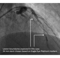
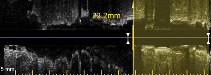
The ADAPT-DES study reported the use of IVUS was associated with a choice of longer stents. 2 With IVUS, you can confirm “healthy-to-healthy” landing zones by checking the plaque burden and tissue type at the lesion boundaries.
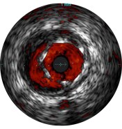
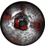
Malapposition is identified by blood behind the stent struts. ChromaFlo imaging colors blood flow red for easy recognition of malapposition and other lumen features.
The incidence of edge dissection after DES implantation is reported to be 10%, with almost 40% of those undetected by angiography. A dissection angle ≥60° or MLA
Stent expansion is a predictor of stent thrombosis and restenosis. Target minimum stent areas post-PCI may include: ≥80% of the average reference lumen areas, 6 mm 3 for DES in non-LM vessels, or other criteria depending on the type of PCI. IVUS helps document your result. 3
Philips has a comprehensive suite of imaging devices and modalities to help you see clearly so you can quickly and easily assess lesion morphology with additional imaging modalities, giving you advanced insights. Experience the benefits from Eagle Eye Platinum digital IVUS catheters.
• Soft, tapered tip with lowest available entry profile and choice of two lengths 4 • GlyDx hydrophilic coating • Long rapid exchange lumen for pushability • Radial access appropriate; fits through all 5F guides 5
• Three radiopaque markers not offered by other IVUS catheters • 10 mm spacing facilitates length estimation without a pullback device or marker wire
• Easy assessment of stent apposition, lumen size and more by highlighting blood flow red at the touch of a button • Provides advanced imaging on the IntraSight interventional applications platform with IVUS Co-registration*


Experience the advanced imaging insights from Philips ChromaFlo and IVUS Co-registration and improve your treatment strategies.
Imaging, physiology, co-registration* and software come together to simplify complex interventions, speed routine procedures and provide improved patient care.
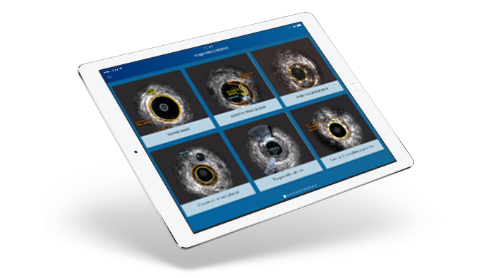
Philips ELIITE Academy is focused on delivering high value and real-time strategic educational programs that meet the evolving needs of our customers. For more information on the available courses, please visit www.igtdacademy.philips.com .


*Co-registration tools available within IntraSight 7 configuration via SyncVision 1. Shlofmitz et al. IMPROVE trial: Study design and rationale. AHJ; (2020) Oct, Vol 228, doi.org/10.1016/j.ahj.2020.08.002 2. Witzenbichler B et al. Relationship Between Intravascular Ultrasound Guidance and Clinical Outcomes After Drug-Eluting Stents: The ADAPT-DES Study. Circulation 2014 Jan: 129,4;463-470. 3. McDaniel M. et al. Contemporary Clinical Applications of Coronary Intravascular Ultrasound. JACC: Cardiovascular Interventions. 2011;4 (11): 1155-67. Liu X et al. A Volumetric Intravascular Ultrasound Comparison of Early Drug-Eluting Stent Thrombosis Versus Restenosis. JACC Cardiovasc Interv. 2009;2:428-34 4. 0.019” entry profile, data on file; tip to transducer lengths offered include 2.5 mm and 10 mm 5. Fits through guide catheters with inner diameters as low as 0.056”; data on file at Philips
Can we help?By clicking on the link, you will be leaving the official Royal Philips ("Philips") website. Any links to third-party websites that may appear on this site are provided only for your convenience and in no way represent any affiliation or endorsement of the information provided on those linked websites. Philips makes no representations or warranties of any kind with regard to any third-party websites or the information contained therein. I understand
You are about to visit a Philips global content page ContinueBy clicking on the link, you will be leaving the official Royal Philips ("Philips") website. Any links to third-party websites that may appear on this site are provided only for your convenience and in no way represent any affiliation or endorsement of the information provided on those linked websites. Philips makes no representations or warranties of any kind with regard to any third-party websites or the information contained therein. I understand
You are about to visit a Philips global content page Continue